
Keratoconus
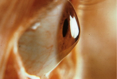
Keratoconus is a progressive eye disease in which the normally round cornea thins and begins to bulge into a cone-like shape. This cone shape deflects light as it enters the eye on its way to the light-sensitive retina, causing distorted vision.
Keratoconus is a non-inflammatory, self-limiting ectasia of the axial portion of the cornea. It is characterized by progressive thinning and steepening of the central cornea. As the cornea steepens and thins, the patient experiences a decrease in vision which can be mild or severe depending on the amount of affected corneal tissue.
Typically, vision loss can be corrected early with spectacles; later, irregular astigmatism requires optical correction with rigid contact lenses. Contact lenses provide a uniform refracting surface and therefore improve vision. Contact lenses can improve vision,however can also scar the cornea. Patients should be informed upon diagnosis that they will likely require contact lenses eventually.
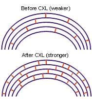
The latest revolutionary Keratoconus treatment is C3R (Corneal Collagen Crosslinking with Riboflavin) that has been proven to strengthen the weak corneal structure & by increasing collagen crosslinking, which are the natural “anchors” within the cornea. These anchors are responsible for preventing the cornea from bulging out and becoming steep and irregular, consequence of advanced Keratoconus.
Causes of Keratoconus:
Keratoconus rarely results in total blindness although it can significantly impair vision and, according to experts, leads to the need for a corneal transplant in up to 20% of cases. While nobody knows the cause of keratoconus, there is evidence that the disease has genetic origins, possibly made worse by environmental factors.It normally affects both eyes, though it typically progresses at different rates.
In most people, keratoconus begins during their teen years and slowly worsens before stabilizing in their 30s or 40s.
Keratoconus is a corneal disease affecting 1 in 2000 individuals. The disease usually starts in young adults in most people, keratoconus begins during their teen years and slowly worsens before stabilizing in their 30s or 40s and is characterized by progressive thinning and outward protrusion of the cornea.
It is normally treated with rigid contact lenses or specialize keratoconus contact lenses which are contoured to address the bulging cornea and to improve vision. With newer treatments like C3R, the progression of the keratoconus can be arrested.
If keratoconus left untreated can result in severe corneal thinning, bulging and scarring of the cornea (corneal hydrops) making the patient blind. The scarred cornea would eventually require a corneal transplant procedure.
Signs & Symptoms
- Frequent changing of glasses or contact lens prescriptions.
- Blurring and distortion of vision.
- Glare and sensitivity to light especially during night Irritation.
- Scarring of the cornea.
Treatment
The early detection of keratoconus is important to arrest the progression of keratoconus and corneal irregularities. In early stages, keratoconus can be diagnosed after a detailed eye examination, which includes Retinoscopy, Slitlamp examination and tests like Topography, pentacam to check the curvature of cornea and Pachymetry to determine corneal thickness.
In advance stages keratoconus, is easily diagnosed on slit lamp and topography. In advanced stages, corrective prescription glasses do not help the patient to see clearly and they have to switch to using hard or semi soft contact lenses. A proper contact lens fit is crucial to obtain adequate vision and wearing comfort. Poorly fit or outdated contact lenses can be uncomfortable and lead to additional complications like corneal abrasions, scarring or infection.
Although there are no medicines known which will prevent progression of the disease, mild cases of keratoconus can be successfully treated with glasses or specially designed contact lens.
The keratoconus changes are first detected on the back surface before they present on the corneal front surface. Thus keratoconus can be detected early before any signs & symptoms by evaluating corneal back surface topography map. Today progressive keratoconus are stablised by a Corneal Collagen Cross Linking (C3R) procedure, INTACS and avoiding Corneal transplant surgery.
COLLAGEN CROSS LINKING:
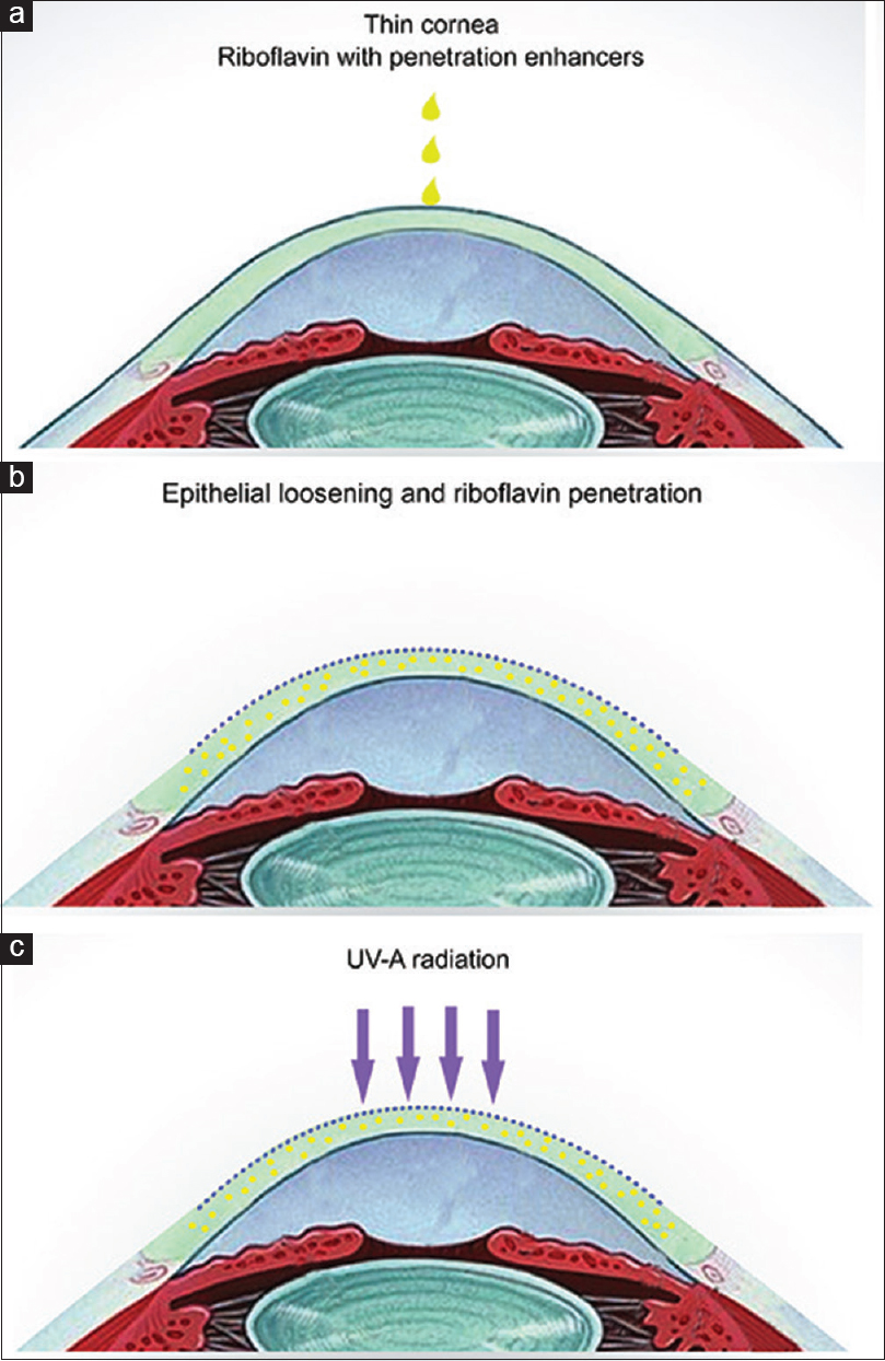
Corneal collagen cross-linking is a technique which uses UV light and a photosensitizer to strengthen chemical bonds in the cornea. The goal of the treatment is to halt progressive and irregular changes in corneal shape known as ectasia. These ectatic changes are typically marked by corneal thinning and an increase in the anterior and/or posterior curvatures of the cornea, and often lead to high levels of myopia and astigmatism. The most common form of ectasia is keratoconus and less often ectasia is seen after laser vision correction such as LASIK
Intacs
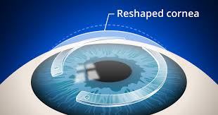 Intacs are used to regularize the shape of the cornea. After the above procedures of intacs and C3R specialized contact lenses or implantable contact lenses can be given to achieve full visual potential. INTACS MADE MORE EASIER,SAFER,AND ACCURATE WHEN DONE BY FEMTOSECOND LASER. The advent of femtosecond laser has made creation of accurate channels for intacs or corneal rings at precise depth and size in the cornea, thus entering the effectiveness after corneal rings to flatten the keratoconus.
Benefits of Intacs for Keratoconus:
- Safe, removable, replaceable.
- Reduces myopia and astigmatism associated with keratoconus.
- Restores the cornea to a more natural dome shape.
- Minimally invasive surgical procedure.
- Recovery period is days, compared to months for a corneal transplant.
- Improves quality of life.
- May delay need for corneal transplant.
Keratoplasty (Corneal Transplantation):
It is the option when not all the above methods help patients to get optimum useful vision. In this procedure the cornea, which forms the transparent outer covering of the eye, is replaced by a donor cornea. It is an intraocular surgery and requires long-term and consistent follow-up.
Keratoconus is a disease that causes a progressive thinning of the cornea (the clear front portion of the eye). As a result of this condition, the normal outward pressure from within the eye causes the cornea to progressively bulge into a cone-like shape.
Specialisation
- Lasik / Refractive Surgeries
- Cornea
- Cataract
- Keratoconus
- Retina Surgery
- Glaucoma Services
- Oculoplastic Surgery
Contact Information
Dr. Shaila Rajkumar Patel
MBBS, DNB Ophthal (Arvind Eye Hospital), F.C.R.S. (Fellowship in Cornea and Refractive Surgery), Fellowship in Advanced Phaco, MNAMS
301, 3rd Floor, MTNL Road, Near Don Bosco School, Mira Road (East), Thane - 401 107
+91 83698 81371